1
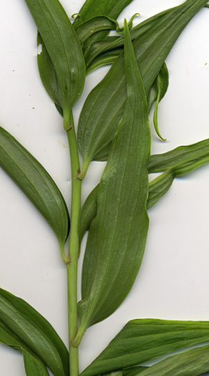
The picture on the left shows the leaf deformity, but the chlorosis is hard to see.
If you click the picture, the full view will show a fairly uniform sprinkling of spores on
the tops of the leaves and on the stem.
While there are few spores on the leaf bottoms, there is some spore clustering.
The leaf junctions are reddish in color but there are no spores there as on all the trees.
That was unexpected.
One interesting white canker clue is that there always seems to be spores
clustering on the underside of the leaf just about a half-inch after the leaf widens.
Coincidence? I flipped this sample over and scanned the other side.
The same pattern of clustering appeared, so I assume this lily IS susceptible to white canker.
2
But the biggest surprises came through microscopic exams of parts of the plant.
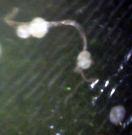
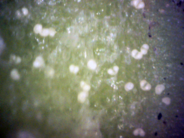
The picture above shows several spores with their typical shape and a small stalk
(lower right corner), just as we've seen on trees and shrubs.
It also shows two of the spores connected by a hypha, which is embedded
in the leaf, proving that these spores can infect a lily.
Note that the spore seems to change shape as it germinates to send out a hypha that in turn creates more spores. (400x)
5
My biggest surprise came when I ripped off a leaf and microscopically examined the leaf scar.
To my amazement I saw spores there!
I had previously assumed they only grew on the surface of stems and leaves,
and that only hyphae grew inside the tissue.
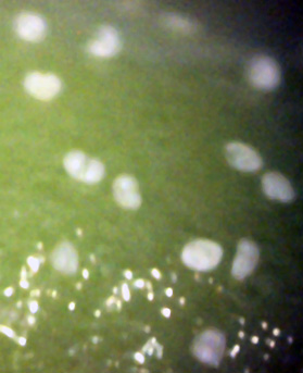
The pictures above show several spores within the scar area.
This scar area was exposed only a minute before this picture was taken,
so it is unlikely they spread to this area and instantly grew in that time period. (400x)

6
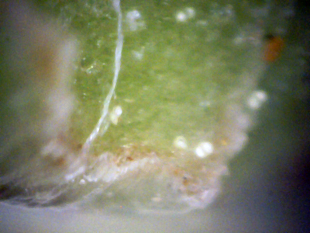
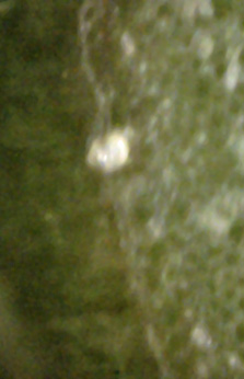
This view is somewhat unusual since it shows a side view of several spores which
are on the edge of a leaf. (400x)
8
Having now found evidence that these spores can grow within a leaf scar,
I wondered if they could also grow within the stem.
Fortunately, this being a plant, it was easy to make a clean cut of the stem with a razor blade.
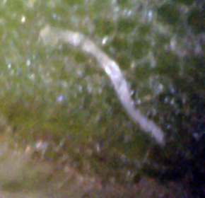
Sure enough, I saw white spores within this cross cut. The picture above shows one of them connected to a hypha on the cut surface.
7
This picture shows a cut hypha pushed out of the cut stem.
Apparently the internal pressure forced it out, since the cut took place only a minute earlier. (400x)
9
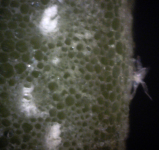
This picture seems to show white spores buried just under the cut surface,
and a hypha star (mycelium) on the stem skin. (400x)
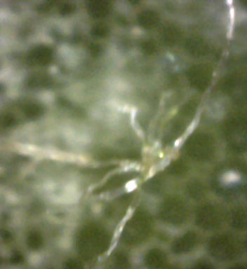
10
A buried hyphae star, or mycelium on the stem's cut surface.
These structures must be pretty tough to survive a razor cut fairly intact, and then pop out!
(400x)
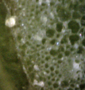
11
This picture shows several buried spores, one of which is actually
penetrating the stem's skin. (400x)

13
Here we have spores on the edge of the cross-cut - the skin of the stem.
The rightmost spore apparently hasn't had time to do anything yet.
The one in the middle is losing its yellow color as it starts to send out hypha.
The spore on the left apparently has been cut, and its jelly-like insides have gushed out in blobs.
The two spores on the right seem to be transforming themselves into spore generators by changing their color and shape.
(400x)

14
This is a side view of a spore just below the surface.
To its right is a mass of a white jelly-like substance just below the skin.
This probably is the cause of the chlorosis seen on the plant - visible white canker. (400x)
12
↓
Here we have a normal white spore on the right.
On the left it appears as if a white spore is giving birth to a yellow/new spore (red arrow). (400x)
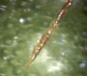
15
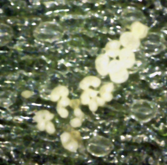
Above is a picture of a rare mottled brown hypha sticking up from the stem crosscut.
It extends about the same distance upward again, and then ends in an abrupt cut,
due to the razor. It must have been under extreme pressure to pop up so high. (400x)
16
↓
←
←
↑
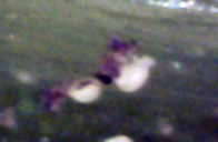
Here again is a lower leaf surface with lots of white spores,
some apparently giving birth to yellow spores.
Several leaf stomata (breathing pores) are also shown (orange arrows).
One yellow spore is developing a cleft and turning into a mature spore (blue arrow).
The bottom yellow spore (pink arrow) is likely a child of the parent white spore next to it. (400x)
17
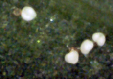
I've always wondered what''s inside those white spores.
This spore apparently has broken, spilling its insides (brown mess). (400x)
18
↑
↓
Here we apparently have two spores in the process of making new spores.
The parent spores are white. The new spores are the small yellow globules budding to their side. (400x)
Some of the most beautiful flowers in our yard are produced by a cluster of Asiatic lilies.
I noticed that they were suffering from chlorosis (yellowing), some of the leaves were a bit curled,
and the new leaves were pretty deformed.
This is a classic symptom of white canker. So I cut the top of a plant off for analysis.
As an experiment, I took one leaf that had a good sprinkling of white
mature spores on the upper surface, washed it under tap water for a few seconds,
and then gently rubbed it dry for a few seconds.
When I scanned it again, 99% of the spores were missing, indicating that they
weren't attached to the top leaf surface very firmly.
When I took another leaf and tried gently scraping its leaf surface with a razor blade,
I was unable to remove any spores.
I'm not sure why I couldn't remove any spores (maybe the blade only hit the high points on the leaf?).
When I started analyzing this lily, I thought I wouldn't learn much from it,
and almost didn't try. But to my surprise, it had a lot to teach me. Specifically,
- The white spores seem to be generators of the yellow spores, which often grow to the side of them until they are full size. When working with these diseased plants for awhile, I noticed that my hands had numerous yellow spores on them, while there were no white spores on them.
- You can easily wash the white spores off the leaf surface.
- The interior of the leaves and stem are loaded with spores and hyphae.
- The loss of greening on the leaves only appears to be chlorosis, but is actually caused by a buildup of a spore-related substance and hyphae. Microscopic analysis shows the leaf cells themselves keep their color.
